Noninvasive bone fracture risk assessment using Raman spectroscopy
Osteoporosis is a worldwide concern that causes millions of fractures per year which may lead to mortality or morbidity. Women over the age of 40 are twice as likely to be osteoporotic than men at the same age. This bone disease is characterized by reduced bone mass and low bone mineral density.
Currently, osteoporosis is diagnosed by measuring bone mineral density (BMD) using dual-energy X-ray absorptiometry (DXA). Despite BMD being correlated with bone strength, it is a poor predictor of fracture risk. The bone’s tissue mechanics may be a more accurate predictor for bone fragility or fracture risk, but it is not clinically feasible to assess these properties.
Our lab’s goal is to use Raman spectroscopy as a pre-clinical, non-contact optical assessment of bone fragility and fracture risk. Raman spectroscopy is a non-destructive vibrational spectroscopy technique that can reveal important biochemical information about the bone mineralization and matrix. We aim to study biochemical changes that occur in different mouse models for diagnosis in mice and predict the biomechanical changes.

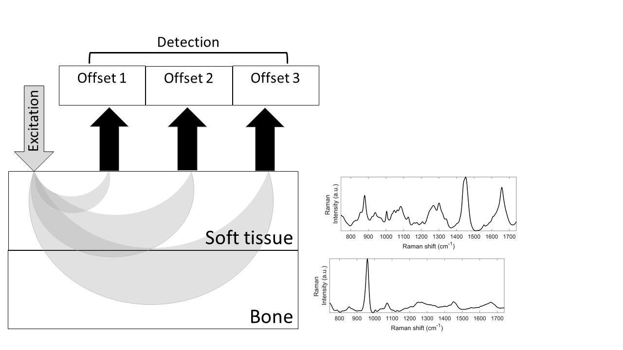
By implementing spatially offset Raman spectroscopy, we are capable of performing measurements in vivo. Our detected signal will be a mixture of signals from the soft tissue and the bone. We have developed algorithms to extract the bone’s spectrum which improves our in vivo bone quality assessment using Raman spectroscopy.
Future directions will be scaling up murine tibia experiments to human finger bone quality assessment.
Collaborators: Hani Awad